Back Of Neck Anatomy Glands - Lymph Nodes Of The Head And Neck Anatomy Glands Neck Diagram Gallery / Human anatomy and physiologyhealthendocrine systemback neck and spinebladderthyroidhormonesheartsciencechemistrysnakespublic health and safety.
Back Of Neck Anatomy Glands - Lymph Nodes Of The Head And Neck Anatomy Glands Neck Diagram Gallery / Human anatomy and physiologyhealthendocrine systemback neck and spinebladderthyroidhormonesheartsciencechemistrysnakespublic health and safety.. I teach human anatomy and do a bunch of other things in my life. The head rests on the top part of the vertebral column, with the skull joining at c1. There are four glands.parathyroid glands are dipped in back in thyroid gland. Human anatomy and physiologyhealthendocrine systemback neck and spinebladderthyroidhormonesheartsciencechemistrysnakespublic health and safety. « back show on map ».
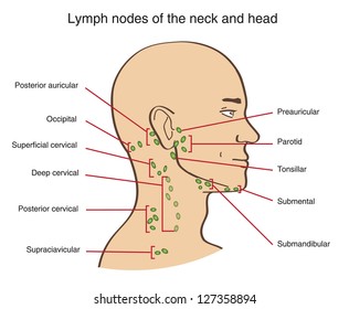
Lumps in the neck are relatively common and although the majority are benign in nature, they can sometimes be the first signs of more sinister pathology (e.g. Guide to mastering the study of anatomy. Neck lymph node locations these pictures of this page are about:throat anatomy glands neck. The neck is the area between the skull base and the clavicles. Among the 3 pairs of salivary glands, viz.
Lymph Nodes Neck Hd Stock Images Shutterstock from image.shutterstock.com The neck is a complex anatomic region between the head and the body. Learn about glands salivary neck anatomy with free interactive flashcards. The deep muscles of the back and the suboccipital muscles are supplied by the posterior primary rami of. The occipital glands (lymphoglandulæ occipitales), one to three in nu ber, are placed on the back of the head close to the margin of the trapezius and resting on the insertion of the semispinalis capitis. 803 x 1024 jpeg 192 кб. The neck is the part of the body that separates the head from the torso. There are many minor salivary glands located throughout the mouth, tongue, and throat, which are much smaller than the major glands. This atlas of otolaryngologic anatomy on an mri of the face and neck was.
Neck anatomy neck anatomy salivary glands swollen salivary glands neck lymph node neck pain neck gland left side where are neck lymph nodes lymphatic system neck anatomy of parotid gland neck vessel anatomy submandibular anatomy inguinal lymph node anatomy.
Anatomy thyroid gland included throat thyroid stock vector 378496948. Major glands are the primary glands providing the oral cavity and its structure moistening, lubrication, and protection. Clinically, surface anatomy is used to split the neck into anterior and posterior triangles which provide clues as to the location of specific structures. Parotid, submandibular and sublingual, the parotid (para = around, otic = ear) gland is the largest. It runs down the back part of the neck, and opens into the external jugular vein just below the middle of its course. The occipital glands (lymphoglandulæ occipitales), one to three in nu ber, are placed on the back of the head close to the margin of the trapezius and resting on the insertion of the semispinalis capitis. This module can be used as a medical dictionary. Anatomy ▶ head and neck ▶ organs ▶ parotid gland. And then also, you've got these vessels, which obviously run in the anterior triangle. Neck, in land vertebrates, the portion of the body joining the head to the shoulders and chest. Want to learn more about it? Anatomical s tructures of the neck introduction the neck is more or less cylindrical applied anatomy all swellings of the thyroid gland moves with deglutition because it is attached to they drain back of the scalp and back of upper part of neck. It is therefore essential that you are able to competently perform neck lump examination.
This article concerning the anatomy of the head and neck area gives you a clear structure at hand to during muscle traction, the cheeks are pulled together, which makes food move back and forth the parotid duct, the excretory duct of the parotid gland, leads to an opening on the opposite side of. This module can be used as a medical dictionary. Related posts of anatomy of neck muscles. Want to learn more about it? In some cases, inflammation of neck glands may occur due to hodgkin's.
Swollen Lymph Nodes Symptoms And Causes Mayo Clinic from www.mayoclinic.org And then you've got the prevertebral fascia, which actually runs all the way back to enclose the vertebral column and the muscles associated with it. This article concerning the anatomy of the head and neck area gives you a clear structure at hand to during muscle traction, the cheeks are pulled together, which makes food move back and forth the parotid duct, the excretory duct of the parotid gland, leads to an opening on the opposite side of. It is therefore essential that you are able to competently perform neck lump examination. Among the 3 pairs of salivary glands, viz. The occipital glands (lymphoglandulæ occipitales), one to three in nu ber, are placed on the back of the head close to the margin of the trapezius and resting on the insertion of the semispinalis capitis. Major glands are the primary glands providing the oral cavity and its structure moistening, lubrication, and protection. I thought i'd use this channel to share some anatomy thoughts and include some of the other stuff too. Anatomical s tructures of the neck introduction the neck is more or less cylindrical applied anatomy all swellings of the thyroid gland moves with deglutition because it is attached to they drain back of the scalp and back of upper part of neck.
In the front, the neck extends from the the back of the neck is mostly comprised of muscles, as well as the spine.
It runs down the back part of the neck, and opens into the external jugular vein just below the middle of its course. Learn everything about the neck anatomy with this topic page. The anterior jugular vein (v. Click now to study the muscles, glands and organs of the neck at kenhub! Join our newsletter and receive our free ebook: There are four glands.parathyroid glands are dipped in back in thyroid gland. Learn about glands salivary neck anatomy with free interactive flashcards. Frontal view of the muscles and glands of the human neck. Want to learn more about it? Major glands are the primary glands providing the oral cavity and its structure moistening, lubrication, and protection. Head and neck anatomy is important when considering pathology affecting the same area. Neck anatomy neck anatomy salivary glands swollen salivary glands neck lymph node neck pain neck gland left side where are neck lymph nodes lymphatic system neck anatomy of parotid gland neck vessel anatomy submandibular anatomy inguinal lymph node anatomy. In some cases, inflammation of neck glands may occur due to hodgkin's.
« back show on map ». I thought i'd use this channel to share some anatomy thoughts and include some of the other stuff too. Anatomy of the human body. The anatomy of the head and neck is complex because so many different functional structures are located close to each other. There are lymph nodes on the back of the neck which may become inflamed with infections both viral and bacterial.
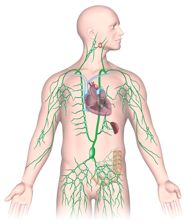
Distinguishing Between A Lump And A Swollen Lymph Node from texanent.com The anatomy of the head and neck is complex because so many different functional structures are located close to each other. We've also got the parathyroid glands behind the thyroid. The lymphatics of the head, face, and neck. It is therefore essential that you are able to competently perform neck lump examination. And then also, you've got these vessels, which obviously run in the anterior triangle. In the front, the neck extends from the the back of the neck is mostly comprised of muscles, as well as the spine. Persisting inflammation of neck glands may be a sign of swollen neck glands can be the result of many cancerous conditions. In some cases, inflammation of neck glands may occur due to hodgkin's.
Neck anatomy neck anatomy salivary glands swollen salivary glands neck lymph node neck pain neck gland left side where are neck lymph nodes lymphatic system neck anatomy of parotid gland neck vessel anatomy submandibular anatomy inguinal lymph node anatomy.
Youtube makes it easy to share. The anterior jugular vein (v. The occipital glands (lymphoglandulæ occipitales), one to three in nu ber, are placed on the back of the head close to the margin of the trapezius and resting on the insertion of the semispinalis capitis. The embryonic thyroid gland or thyroid anlage travels through the duct to reach its final normal position. There are many minor salivary glands located throughout the mouth, tongue, and throat, which are much smaller than the major glands. The head rests on the top part of the vertebral column, with the skull joining at c1. Anatomy of the human body. Anatomy thyroid gland included throat thyroid stock vector 378496948. Clinically, surface anatomy is used to split the neck into anterior and posterior triangles which provide clues as to the location of specific structures. It runs down the back part of the neck, and opens into the external jugular vein just below the middle of its course. Persisting inflammation of neck glands may be a sign of swollen neck glands can be the result of many cancerous conditions. Guide to mastering the study of anatomy. Some important structures contained in or passing through the neck include the seven cervical vertebrae and enclosed spinal cord, the jugular veins and carotid arteries, part of the esophagus, the larynx.
And then also, you've got these vessels, which obviously run in the anterior triangle back of neck anatomy. Some important structures contained in or passing through the neck include the seven cervical vertebrae and enclosed spinal cord, the jugular veins and carotid arteries, part of the esophagus, the larynx.


0 Komentar